Visual Representation

Eukaryotic animal cell coloring nucleolus black – A clear visual representation of a eukaryotic animal cell, particularly highlighting the nucleolus, is crucial for understanding its function within the cell’s overall structure. This aids in comprehension of the nucleolus’s role in ribosome biogenesis and other vital cellular processes. A well-designed diagram simplifies complex cellular architecture, making it accessible to a wider audience.
The following diagram uses a simplified representation to emphasize the nucleolus’s location and appearance within the nucleus. It omits many cellular components for clarity, focusing solely on the key structures relevant to this discussion.
Eukaryotic Animal Cell Diagram with Highlighted Nucleolus
|
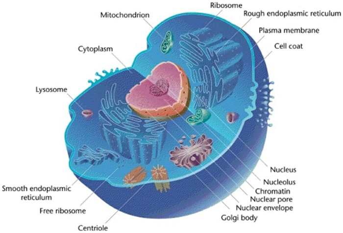
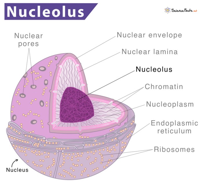
Imagine a circle representing the cell membrane. Within this, a larger, slightly less defined circle represents the nucleus. Inside the nucleus, a smaller, intensely dark-colored (black) circle is the nucleolus. The cytoplasm, the area outside the nucleus, should be depicted with a lighter shading, suggesting a less dense area compared to the nucleus. Various other organelles, such as mitochondria (small oval shapes) and the endoplasmic reticulum (a network of interconnected tubes and sacs), can be represented schematically, but are not the focus. The overall structure should convey a sense of the relative sizes and locations of the organelles, with the black nucleolus standing out prominently within the nucleus. |
Caption: This diagram illustrates a simplified eukaryotic animal cell. The cell membrane encloses the cytoplasm containing various organelles. The nucleus, a large, membrane-bound organelle, houses the genetic material (DNA) and contains a prominent, darkly stained structure called the nucleolus (shown in black). The nucleolus is the site of ribosome biogenesis, a crucial process for protein synthesis within the cell. The diagram emphasizes the location and visual prominence of the nucleolus within the cellular architecture. |
Comparison with Other Cell Structures

The nucleolus, a prominent structure within the nucleus, plays a crucial role in ribosome biogenesis. Understanding its characteristics and comparing it to other cellular components helps clarify its unique function within the complex machinery of the eukaryotic animal cell. This comparison will highlight size, location, and functional differences and similarities between the nucleolus and other organelles, including the nucleus itself, and will also address the differences and similarities between nucleoli in animal and plant cells.The nucleolus, typically spherical and relatively large compared to other nuclear structures, is centrally located within the nucleus.
Its size varies depending on the cell’s metabolic activity; cells actively synthesizing proteins tend to have larger nucleoli. In contrast, other organelles such as mitochondria, which are involved in energy production, are generally smaller and distributed throughout the cytoplasm. Lysosomes, responsible for waste degradation, are also smaller and scattered throughout the cytoplasm. The endoplasmic reticulum, a vast network involved in protein and lipid synthesis, is extensive and occupies a significant portion of the cytoplasm, a vastly different distribution compared to the nucleolus’s confined location within the nucleus.
The Golgi apparatus, involved in protein modification and packaging, is also a relatively large organelle compared to the nucleolus but differs in its structure and function.
Nucleolus versus Nucleus
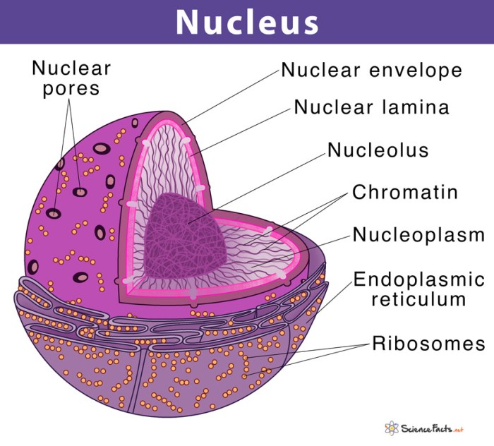
The nucleolus is a distinct sub-compartment
within* the nucleus. The nucleus itself houses the cell’s genetic material (DNA), organized into chromosomes, and is enclosed by a double membrane, the nuclear envelope. The nucleolus, lacking a membrane, is not a separate organelle in the same way as mitochondria or lysosomes are. While the nucleus controls the cell’s overall activity and contains the genetic blueprint, the nucleolus is focused on a specific task
ribosome production. The nucleus’s functions are far broader, encompassing DNA replication, transcription, and regulation of gene expression, whereas the nucleolus’s function is much more specialized.
Nucleolus in Animal and Plant Cells
While the fundamental function of the nucleolus – ribosome biogenesis – remains the same in both animal and plant cells, there might be subtle differences in size and organization. Plant cells, due to their larger size and often higher metabolic activity related to photosynthesis, may exhibit larger nucleoli than some animal cells. However, this is not a universal rule and depends on the specific cell type and its metabolic demands.
Focusing on the intricacies of a eukaryotic animal cell, remember to color the nucleolus black for accurate representation. This detail is crucial for understanding cellular structure, much like the careful coloring of the animals found in a desert oasis is important for a complete picture; you can find some great examples at desert oasis animals coloring pages.
Returning to the cell, the contrast of the black nucleolus against other cellular components aids in visualization and understanding.
The structural organization of the nucleolus might also exhibit some minor variations, although the core process of ribosome assembly remains conserved across both plant and animal kingdoms. The differences are generally minor compared to the significant functional similarities.
Nucleolar Dysfunction and Disease: Eukaryotic Animal Cell Coloring Nucleolus Black

The nucleolus, despite its seemingly simple structure, plays a crucial role in ribosome biogenesis and cell function. Disruptions to its normal activity, termed nucleolar dysfunction, can have profound consequences for the cell and ultimately contribute to the development of various diseases. This dysfunction manifests in a variety of ways, impacting cellular processes from protein synthesis to stress response.Nucleolar dysfunction arises from a multitude of factors, including genetic mutations, environmental stressors, and viral infections.
These factors can disrupt the intricate balance of the nucleolar processes, leading to a cascade of downstream effects that negatively impact cellular health and potentially contribute to disease pathogenesis.
Consequences of Nucleolar Dysfunction
Disrupted nucleolar function leads to a reduction in ribosome production, directly impacting protein synthesis. This deficiency can compromise the cell’s ability to perform its essential functions, leading to cellular stress, apoptosis (programmed cell death), and ultimately, tissue damage. Beyond reduced ribosome biogenesis, nucleolar dysfunction can also affect the cell’s response to stress, impairing its ability to cope with various insults.
This compromised stress response further exacerbates cellular damage and contributes to disease progression. Furthermore, nucleolar dysfunction can alter the expression of specific genes, contributing to the development of abnormal cellular phenotypes.
Diseases Linked to Nucleolar Abnormalities
Several diseases have been linked to nucleolar dysfunction. For example, various cancers exhibit altered nucleolar morphology and function. The nucleolus often displays enlargement and increased numbers of nucleolar organizer regions (NORs) in cancer cells, reflecting its role in supporting the high rate of protein synthesis required for rapid cell proliferation. Additionally, several ribosomopathies, a group of disorders caused by defects in ribosome biogenesis, are directly associated with nucleolar abnormalities.
These include Diamond-Blackfan anemia, characterized by bone marrow failure and reduced red blood cell production, and Treacher Collins syndrome, a condition affecting craniofacial development. These conditions highlight the critical role of the nucleolus in maintaining normal cellular function and development.
Mechanisms Linking Nucleolar Dysfunction to Disease
The mechanisms linking nucleolar dysfunction to disease are complex and multifaceted, often involving a combination of factors. Genetic mutations affecting genes involved in ribosome biogenesis can directly disrupt nucleolar function, leading to the characteristic features of ribosomopathies. For instance, mutations in ribosomal protein genes are frequently observed in Diamond-Blackfan anemia. Furthermore, environmental stressors, such as exposure to certain toxins or infections, can indirectly impair nucleolar function by inducing cellular stress and activating pathways that interfere with ribosome biogenesis.
This disruption of the delicate balance within the nucleolus ultimately leads to compromised cellular function and contributes to the development of disease. The altered gene expression patterns observed in nucleolar dysfunction can further exacerbate these effects, creating a complex interplay of factors that drive disease pathogenesis.
Further Research and Exploration
The nucleolus, despite its seemingly simple structure, remains a fascinating and vital organelle ripe for further investigation. Its central role in ribosome biogenesis and its increasingly recognized involvement in diverse cellular processes necessitate continued research to fully elucidate its complexities and potential therapeutic applications. Unraveling the intricacies of nucleolar function holds immense promise for advancing our understanding of both normal cellular physiology and disease pathogenesis.The multifaceted nature of the nucleolus presents numerous avenues for future research.
A deeper understanding of the dynamic interplay between the nucleolus and other cellular compartments, particularly the cytoplasm and the endoplasmic reticulum, is crucial. Further investigation is also needed into the precise mechanisms regulating nucleolar size and structure in response to various cellular stresses and stimuli. This includes exploring the role of post-translational modifications of nucleolar proteins and the impact of environmental factors on nucleolar function.
Nucleolar Research Applications in Medicine
Research into nucleolar function has direct implications for the development of novel therapeutic strategies. Dysregulation of nucleolar function is implicated in a wide range of diseases, including cancer, neurodegenerative disorders, and viral infections. Understanding the molecular mechanisms underlying these dysfunctions could lead to the development of targeted therapies aimed at restoring normal nucleolar activity or inhibiting its aberrant function in disease states.
For example, identifying specific nucleolar proteins crucial for cancer cell proliferation could lead to the development of novel anti-cancer drugs that selectively target these proteins. Similarly, manipulating nucleolar function could offer new avenues for treating viral infections by interfering with viral replication processes that rely on nucleolar components.
Advanced Imaging Techniques and Nucleolar Function, Eukaryotic animal cell coloring nucleolus black
Advanced microscopy techniques are revolutionizing our understanding of cellular structures and processes, and the nucleolus is no exception. Super-resolution microscopy, such as stimulated emission depletion (STED) microscopy and photoactivated localization microscopy (PALM), allows for visualization of nucleolar substructures with unprecedented detail, revealing the intricate organization of nucleolar components and their dynamic interactions. These techniques can provide insights into the spatial organization of ribosomal RNA transcription, processing, and assembly, and how these processes are affected by cellular stress or disease.
Furthermore, live-cell imaging techniques, coupled with fluorescently tagged nucleolar proteins, allow for the real-time observation of nucleolar dynamics, providing crucial information on the temporal regulation of nucleolar function. For instance, using time-lapse microscopy with fluorescently tagged ribosomal proteins, researchers could observe the movement and assembly of ribosomes within the nucleolus, providing a detailed understanding of ribosome biogenesis. This detailed visualization could reveal subtle changes in nucleolar structure and function not detectable with conventional microscopy techniques, leading to a more comprehensive understanding of nucleolar involvement in various biological processes and disease states.
